2020年7月28日 星期二

(新)眼皮 割雙眼皮 眼皮整形 縫雙眼皮 雙眼皮手術 公告:請先矯正屈光不正.再進行雙眼皮手術.萬分感激.

 (新)眼皮 割雙眼皮 眼皮整形 縫雙眼皮 雙眼皮手術 公告:請先矯正屈光不正.再進行雙眼皮手術.萬分感激.

公告:請先 矯正屈光不正 .再進行雙眼皮手術.萬分感激.


anatomy-physiology-of-eyelids-15-638.jpg


「 眉目傳情」,這是描寫美麗動人心靈之窗的眼睛,所表現出的的各種情感活動。然而什麼樣的眼睛才算美呢?或許有人會說一雙炯炯有神的大眼睛,雙眼皮再配上墨黑的雙眉這就是美。

其實,這還不足夠,尚須與面部其他器官(如臉形、鼻形、內外眼角間距、嘴形)協調一致,才較為理想。


雙眼皮類型

封閉式(內雙)                              半開放式 (東方))                         全開放式 (西方)

雙眼皮種類105-09-05. - 複製.jpgeyelid classification - 複製.jpgcgm9564e7a92b2ffb17201b12592add2c45482.jpg


雙眼皮手術成功的標準


  雙眼皮的手術成功,因為個人的審美觀點不同,很難一致認定。
  但雙眼皮看起來自然、對稱則是成功的基本標準。術後雖有刀痕,但因隱藏在雙眼皮皺摺內,故睜眼時是看不到的。


  不宜做雙眼皮


  .兩眼間距離近的人,手術後效果較不理想。
  .圓形眼的人此時需合併其他手術(隆鼻術、內眥贅皮整形)來改善。
  .有嚴重內科疾病。
  .眼睛患眼疾時,暫不宜做雙眼皮手術。


手術方式 : 依年紀及眼皮鬆弛程度,可分為下列:

  雙眼皮縫合手術(縫合式雙眼皮手術) 雙眼皮切割手術 (切開式雙眼皮手術)
原理 兩眼各 5 針皮下埋線方式。 切除 上眼皮多餘的脂肪 同時可將多餘的眼皮脂肪做切除 。
優點 手術時間短、傷口小、恢復期短、消腫快、術後不必拆線、手術 滿 3 天後就不太會腫。 持久的眼皮手術 。
缺點 雙眼皮鬆開的機會比較高,看縫合技巧而定,也可維持永久。  2-3 年後約有 2 成的人會因縫線被吸收而恢復原狀。 切開的皮膚會留下疤痕且浮腫期較長。   手術後 1 個月,看起來比較自然。
適用對象      眼皮較薄、皮下脂肪少、眉眼間距較短者。      眼睛較浮腫、眼皮較厚、眼皮下垂、眼皮脂肪過多、睫毛倒插、上眼皮的皮膚鬆弛或過多,去除不需要的皮膚或脂肪。
手術時間 約 40 分    約 1.5 小時
麻 醉 局部麻醉 局部麻醉
復原時間 約 5-7 天,愈年輕復原速度愈快    1 週消腫,但 約 1-2 月才會
術後照顧   
    42小時內冰敷數次(1次15-30 分鐘),3天後改溫敷,以利退瘀消腫。
•    以無菌棉棒沾生理食鹽水清潔,塗抹消炎藥膏避免感染。
•    避免菸、酒、辣椒、等刺激性食物。
•    避免長時間視線往下閱讀或打電腦。
•    不需拆線。
   

請依照醫護人員指示回診,並按時服藥、冰敷、避免搬重物、低頭及俯臥趴睡等 。

術後照顧同左。


•    1 週後拆線,拆線後回家可洗臉碰水,但不可用力去揉。局部可擦上抗疤凝膠,或眼尾會貼上膚色透氣紙膠至少二個月,進行疤痕護理。

請詳閱完上列內容以及內文之超連結,謝謝。

下列為諮詢影片,僅供參考。


2019年4月23日 星期二

Upper Blepharoplasty

Upper Blepharoplasty

JAMA. 2019;321(13):1320. doi:10.1001/jama.2019.1957
Image description not available.
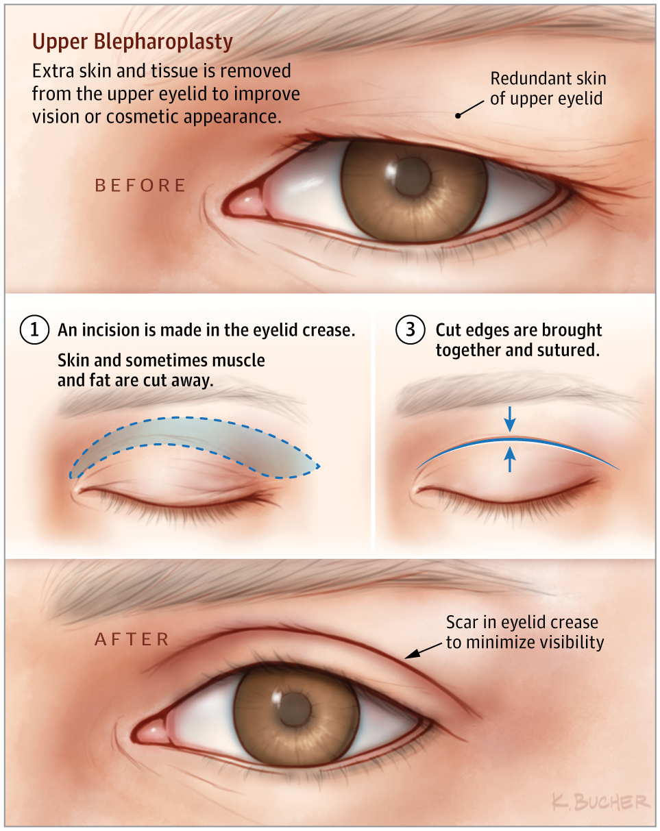
Upper blepharoplasty is a procedure that involves resection of redundant skin and/or musculature of the upper eyelid.
Aging of the Upper Eyelid/Brow Complex
With aging, a patient may notice the upper lids feel “heavy.” This can be caused by the eyebrow descending, upper eyelid skin excess, or both. Upper blepharoplasty, or eyelid lift, is distinct from a brow lift, which elevates the eyebrow. These procedures may sometimes be done together.
Reasons for undergoing an upper blepharoplasty may be purely cosmetic (to reduce the appearance of aging), functional (to reduce obstruction of vision by overhanging skin), or both. Functional upper blepharoplasty involves reduction of the upper eyelid to improve visual obstruction caused by redundant lid tissue. Patients may seek cosmetic upper blepharoplasty with aesthetic concerns regarding overhanging skin, which may cause a less youthful appearance. Clinical evaluation and management of both functional and cosmetic concerns is important. In addition, any ptosis of the upper eyelid (meaning that the margin of the eyelid sits too low and covers the pupil) should be noted. This is caused by poor function of the muscles of the eyelid and is treated differently (ptosis repair).
Patients with eye disease such as glaucoma or dry eye are not candidates for eyelid surgery except in special circumstances.
Basics of Upper Blepharoplasty
The procedure may be done with local or general anesthesia. Technically, the procedure involves identifying the upper eyelid crease on each eyelid and designing a skin excision that removes enough skin to alleviate the issue and preserves enough to allow normal eyelid closure. In some cases, a small amount of the orbicularis oculi (the muscle that closes the eyelid) is removed. Fat is typically preserved over the middle of the eyelid, but some removal may help with aesthetic contour in cosmetic eyelid surgery. When the procedure is complete, the eyelid is closed with sutures. The incision should lie in the eyelid crease, making it less visible after healing.
Recovery
Typically, patients may have mild bruising or swelling around the eyes for 10 to 14 days. Some surgeons may recommend avoidance of makeup for 2 to 3 weeks. Glasses may be worn immediately, but patients may be instructed to avoid contact lenses for a few weeks, depending on surgeons’ preferences.
Complications of blepharoplasty are relatively rare. The primary issue to consider is removal of too much upper eyelid skin, resulting in difficulty closing the eye. This in turn may cause chronic dry eye issues and pose a risk of injury to the cornea. For this reason, conservative skin excision is recommended. Other complications include ptosis of the eyelid (due to injury of the levator muscles), asymmetry of the eyelids, or blindness, which is exceedingly rare.

For More Information

To find this and other JAMA Patient Pages, go to the For Patients collection at jamanetworkpatientpages.com.
The JAMA Patient Page is a public service of JAMA. The information and recommendations appearing on this page are appropriate in most instances, but they are not a substitute for medical diagnosis. For specific information concerning your personal medical condition, JAMA suggests that you consult your physician. This page may be photocopied noncommercially by physicians and other health care professionals to share with patients. To purchase bulk reprints, call 312/464-0776.
Back to top
Article Information
Conflict of Interest Disclosures: None reported.
Source: Weissman JD, Most SP. Upper lid blepharoplasty. Facial Plast Surg. 2013;29(1):16-21. doi:10.1055/s-0033-1333833

2018年8月21日 星期二

Painful Lump in Breast

Painful Lump in Breast

Eventually, the young girl starts to undergo the hormonal changes of becoming a woman and menstruation begins with development of breasts. These changes in the breast tissues will often include itching, pain, and other discomforts, particularly during the menstrual cycle. Sometimes lumps will develop and they can become tender or even painful, but this is actually fairly common and such developments are typically not related to cancer.
Possible Causes of Painful Lump in Breast
  1. Generalized Breast Lumpiness
Once known as fibrocystitis or fibrocystic disease of the breasts, it was eventually realized this condition is so common and it’s, in fact, considered normal. Characterized by many nodular lumps in the breasts or varying areas of thickness in certain areas of the breast, painful lump in the breast can also feel as though there are clusters of small bumps which are tender to touch. There is typically a tendency for this to flare up during the menstrual period or pregnancy and is considered completely normal. Hormone therapy will also exacerbate this condition. Though it can be chronic, it often abates after menopause. With consistent breast exams, you can assure that none of these lumps are cancerous, as they are often benign.
  1. Cysts and Abscess Lumps
  • Cysts are basically sacs of fluid found in soft tissues of the body. Typically, they are only about an inch or two in diameter, if not smaller. They feel rubbery or smooth upon palpation and are atypical in women under the age of 50. When found in the breasts, they can be particularly painful. There is no relation between breast cysts and cancer of the breasts. Physicians can drain or remove cysts.
  • Sebaceous cysts occur at the site of the skin when sebaceous ducts at hair follicles become blocked. The result is an internal sack which swells with sebum, or sebaceous fluid. Though generally not painful, sometimes they can press on certain nerves and become blindingly painful. When such symptoms are caused, physicians will incise the skin and remove the entire cyst to avoid re-growth
  • Abscesses can be a painful situation and always indicate infection. They are one of the ways the body will contain an infection, say, at the site of the breast. This would be an example of painful lump in breast. There may be an accompanying fever with overall illness and malaise. Antibiotics are typically prescribed and surgery is frequently needed to drain the pus from the abscess.
  1. Fat Necrosis
Necrosis means “death of tissue,” so the connotation is generally not a favorable one. However, in this case it would be a potential cause of a painful breast lump. The fat cells in the breasts will become inflamed and altered to form into round lumps which are typically firm upon palpation. The necrosis will typically occur after a firm, bruising impact to the area or some other type of injury in the chest. It may take weeks for fat necrosis to form after an injury occurs.
  1. Adenomas
Another type of lump which will show up in the breasts of younger women is a fibroadenoma. Adenomas are fairly common growths in glandular tissues. They even occur in the testicles of men, where they are entirely more painful. These lumps present as smooth, round, firm, and have defined borders with minimal movement upon palpation. They shift a bit in size with the menstrual cycle and are completely harmless and typically painless.
  1. Blood Clots
The medical name for a blood clot in a vein, which can actually be an immensely painful condition sometimes, is thrombophlebitis, and it will often feel like a firm lump. Typically affecting larger veins crossing the chest from areas under the arms, there will often be redness and pain in the breast area causing painful lump in breast, warmth, with lumpiness along the vein. Such clots are rare, but they should be attended to by a physician.
Could It Be Breast Cancer?
Considering this rather broad question, the general answer is “yes, it could be.” The key word in this answer is, “could.” This means it is a possibility. This is why breast exams first aim to find a lump in the breast.
However, most lumps in the breast, in fact 90 percent found in women between the ages of 20 to 50 years old, are benign. Painful lump in breast usually does not mean breast cancer. It is more often newer, painless lumps that are more cause for concern.
Early signs for real breast cancer include the following:
  • Intractable breast pain
  • Nipple shape changes
  • New lumps which persist even after period changes
  • Discharge from the nipples
  • Swelling and redness, rashes, or other skin irritations at the breast
  • Lumps and swelling at the collarbone or lumps under your arm
  • Any lumps which are hard and have irregular edges
Signs indicative of later breast cancer:
  • Nipple retraction
  • Singular breast enlargement
  • Superficial breast dimpling
  • Lumps which grow larger
  • Skin which presents a texture similar to that of an orange peel
  • Pain in the vaginal area
  • Extreme weight loss, unexplained
  • Armpit lymph nodes which are enlarged
  • Extreme vein visibility on breasts
These symptoms do not mean you have cancer. They mean you need to check with a physician because they are indications of possible breast cancer.
When to See Your Doctor
You should see your doctor if you find any new breast lumps, especially if pain does not reduce after your next period or if a lump which was already present becomes larger or develops irregular edges or other changes. If you notice dimpling, puckering, skin changes, or redness on your breast or breasts, or even nipple inversion, it will be critical to consult a physician. If you see any discharge and you are not pregnant,again, see a doctor.
How Your Breast Normally Feels
Texture of breasts can vary from person to person but generally feels firm to fatty with a slight rope-like consistency with minor nodular consistency. The main tissue is fatty and soft. Breast tissue changes through hormone cycles, often becoming swollen and tender during menstrual cycles. As age progresses, breasts increase in fat and often become larger. When irregular lumps form and painful lump in breast becomes an issue, doctor exams will help ensure the tissue remains healthy and disease is avoided.

2017年4月25日 星期二

Vitreoretinal interface disorders OCT diagnostic indicators

Vitreoretinal interface disorders
OCT diagnostic indicators
Prof.Karim A Raafat-Cairo University

The vitreo-retinal interface has 2 components, the posterior cortical vitreous (collagen type II) and the internal limiting membrane (collagen type IV). Several pathological entities may take place at the Vitreomacular interface including: idiopathic macular hole, lamellar macular hole, Epimacular membrane, Vitreomacular traction syndrome and myopic traction maculopathy (myopic foveoschisis, myopic macular hole without retinal detachment and myopic macular hole retinal detachment). Vitreoretinal interface changes may be encountered in association with diabetic retinopathy (diffuse diabetic macular edema and proliferative diabetic retinopathy). Recently, vitreoretinal interface changes have been incriminated in the pathogenesis of exudative age-related macular degeneration. In addition, certain vitreoretinal disorders may be encountered, that cannot be classified to any of the above, and may represent more than one disorder occurring together. Some of these disorders may be inter-related or represent different stages of the same disorder. Tremendous advances in definition; diagnosis and understanding these disorders have been made after the introduction and evolution of Optical Coherence Tomography (OCT). The pathogenesis of these disorders is still not fully understood and several hypotheses have been speculated: Partial posterior vitreous detachment with persistent vitreo-macular adhesion and traction, splitting of the posterior cortical vitreous (vitreoschisis) with persistent adhesion and traction induced by the outer layer and the internal limiting membrane may act as a scaffold for the proliferation of contractile cells. The rationale of surgical treatment is based on complete relieve of traction on the macular area. In most disorders, this can be achieved by identification, peeling and excision of the posterior cortical vitreous, epimacular membranes and internal limiting membrane. Different stains have been proposed to facilitate visualization and complete peeling of these rather transparent membranes.

Vitreoretinal interface disorders attachment.php?attachmentid=2953&d=1477472735

Vitreoretinal interface disorders attachment.php?attachmentid=2943&d=1477472721

Vitreoretinal interface disorders attachment.php?attachmentid=2942&d=1477472720

Vitreoretinal interface disorders attachment.php?attachmentid=2944&d=1477472724

Vitreoretinal interface disorders attachment.php?attachmentid=2950&d=1477472733

Vitreoretinal interface disorders attachment.php?attachmentid=2947&d=1477472729

Vitreoretinal interface disorders attachment.php?attachmentid=2952&d=1477472735

Vitreoretinal interface disorders attachment.php?attachmentid=2948&d=1477472730

Vitreoretinal interface disorders attachment.php?attachmentid=2949&d=1477472731

Vitreoretinal interface disorders attachment.php?attachmentid=2946&d=1477472726

Vitreoretinal interface disorders attachment.php?attachmentid=2951&d=1477472734

Vitreoretinal interface disorders attachment.php?attachmentid=2941&d=1477472719

Vitreoretinal interface disorders attachment.php?attachmentid=2945&d=1477472725

Source:Piece of ophthalmology

2017年1月26日 星期四

春節假期

公告:春節假期:自106年1月27日下午起至106年2月1日止. 2月2日(大年初六)開始門診.祝新春愉快.闔家平安喜樂.身體健康.

2016年11月30日 星期三

ACEI及ARB、SGLT2抑制劑腎臟保護機轉

[臨床藥學] 報告用大圖 ACEI及ARB、SGLT2抑制劑腎臟保護機轉 (RENOPROTECTION OF ACEIS, ARBS, AND SGLT2 INHIBITORS)

▲腎臟有自我調控機制,第二型糖尿病引起的腎病變破壞了這個機轉。

以前念書時 (對,現在不念書了),老是被考到這個梗:


"為什麼ACEI或ARB可以保護腎臟呢?"

"為什麼可以保護腎臟,又可能造成腎衰竭呢?"

如果你也有一樣的疑問,看這一篇就對了。

"維持自我調控機制是關鍵"

腎臟過濾壓 (filtration pressure),這個壓力受到兩個機轉調控,分別是肌肉血管反應 (myogenic vascular response) 腎小管腎絲球回饋機制 (tubuloglomerular feedback)

肌肉血管反應透過入球、出球小動脈的管徑,以收縮、擴張的差異來調控過濾壓。

腎小管腎絲球回饋機制則是於緻密斑 (macula densa) 感知鈉離子及氯離子的濃度,接著增加腺苷酸 (adenosine) 腎素 (renin) 濃度。

接著就是收縮入球小動脈,降低腎絲球過濾率 (GFR)

糖尿病因為高血糖的關係,近曲小管的SGLT2受體大量回收葡萄糖與鈉,因此降低了緻密斑感受到的鈉離子濃度、抑制腎小管腎絲球回饋機制,最終造成腎絲球過度過濾 (hyperfiltration)

"SGLT2抑制劑恢復失控的自我調控機制"


▲RAS抑制劑與SGLT2抑制劑對腎臟的保護機轉不同。

RAS抑制劑,例如ACEI或ARB,這類藥品擴張出球小動脈的程度"大於"入球小動脈,因此降低腎絲球的壓力,也就是後負荷 (afterload)

而SGLT2抑制劑則是抑制近曲小管的SGLT2受體,增加緻密斑的鈉與氯離子濃度,恢復因為高血糖而失控的自我調控機制。


▲一次比較RAS抑制劑與SGLT2抑制劑的腎臟保護作用機轉。

大圖資料來源:

Nephron Protection in Diabetic Kidney Disease. N Engl J Med 2016; 375:2096-8.

2016年11月25日 星期五

[臨床藥學] 糖尿病治療藥物整理與比較

[臨床藥學] 糖尿病治療藥物整理與比較

Posted: 八月 4, 2015 by 金金金 in Clinical Pharmacy
2
2015-8-4
今天早上參加內科部晨會時,演講的醫師被台下醫師問到 Acarbose是否有什麼肝腎功能不全的使用禁忌,一時之間我心裡也抖了一下、答不上來,於是開啟了今天的藥物資訊,好好把糖尿病用藥做個整理。
還是先公布解答,Acarbose在腎功能 CrCl<25 mL/min的病患為使用禁忌,肝功能禁忌方面仿單未提及,UpToDate則是建議肝硬化(cirrhosis)的病患為使用禁忌,Acarbose作用於腸道,抑制 α-glucosidases和 α-amylase,減少糖分水解成葡萄糖,進而減少葡萄糖吸收,原型藥被吸收進入體內的比例非常低 <2%,但其代謝物會有 35%被吸收,而此代謝物需要靠腎臟排除,因此在腎功能不全的病患會產生蓄積,因此列為禁忌症。
其他藥物的簡單機轉、糖化血色素(HbA1C)降幅、優缺點、禁忌症就如表格不多說囉。
———DPP-4 inhibitors分隔線———
在收納 16,492位有糖尿病及心血管病史或風險的病患、為期2年的 SAVOR trial發現使用 Saxagliptin 比起 placebo 和較高的心衰竭住院(hospitalized for heart failure)風險相關 (HR, 1.27; 95% CI, 1.07-1.51; p=0.007),目前美國 FDA正 review data中。
SAVOR trial:
其他已發表、以心血管事件為終點的 DPP-4 inhibitors臨床試驗方面,Alogliptin (Nesina 耐釋糖)的 EXAMINE trial和 Sitagliptin (Januvia 佳糖維)的 TECOS study則是沒有發現類似相關的風險。
EXAMINE trial & TECOS study:
———SGLT-2  inhibitors分隔線———
目前台灣有的 SGLT 2 inhibitors:Dapagliflozin和 Empagliflozin其腎功能不全的絕對禁忌皆為 eGFR<30 mL/min。而 Dapagliflozin不建議使用在 1.用藥前(baseline) eGFR就已經<60 mL/min的病人 2. 使用後,eGFR一直掉至<60 mL/min的病人。而 Empagliflozin不建議使用在 1.用藥前(baseline) eGFR就已經<45 mL/min的病人 2. 使用後,eGFR一直掉至<45 mL/min的病人。
———Metformin腎功能不全禁忌症分隔線———
2014年11月,TFDA (台灣衛生福利部 食品藥物管理局)公告含metformin成分藥品再評估結果相關事宜:(一)修訂仿單「禁忌症」:腎功能不全患者之「禁忌」內容,修訂為「腎絲球體過濾率(eGFR)小於30 ml/min/1.73 m2禁用」。(二)修訂仿單「用法用量」處:增列「腎絲球體過濾率(eGFR)介於30-45 ml/min/1.73 m2應減量使用」。